Natur und Technik > Mikroskopie
Weitere Unterabschnitte: Biologie | Mikroskopie | Biotechnologie | Chemie | Physik | Physik 2 | Mikrokosmos | Nanokosmos | Wetter und Klima | Weltraum

Chemical Heritage Foundation – Flickr

Curious Expeditions – Flickr

Universal silver microscope

One of two ornate silver microscopes made by George Adams (1709-1773) of Fleet Street, London, for King George III (1738-1820). It is based on the 'Universal Double Microscope', described by Adams in 1746. It is made of brass and steel, covered in beaten silver.Photographed in the Science Museum, London, through its glass case.Use red/cyan anaglyph glasses to see in 3D (red over the left eye)
Bild: kiwizone

Microscopes

The Museum for the History of Science, in Oxford. This is a 3D anaglyph - to view, wear glasses with red on the left eye, and cyan (green-blue) on the right eye.

Fly form Saudi Arabia

This fly specimen, Atherigona bimaculata, was collected at Maraba, Aseer, Saudi Arabia.Image: James Turner, Amgueddfa Cymru.Discover more of these remarkable images on the Up Close With Nature online gallery on 'Rhagor', the collections based website from Amgueddfa Cymru - National Museum Wales.

PROYECTO AGUA** /** WATER PROJECT – Flickr

David (MK) – Flickr

Carl Zeiss Microscopy – Flickr

Carl Zeiss AG, Deutschland

wellcome images – Flickr

St Stev – Flickr

Bild: St Stev

Beetle tracheas

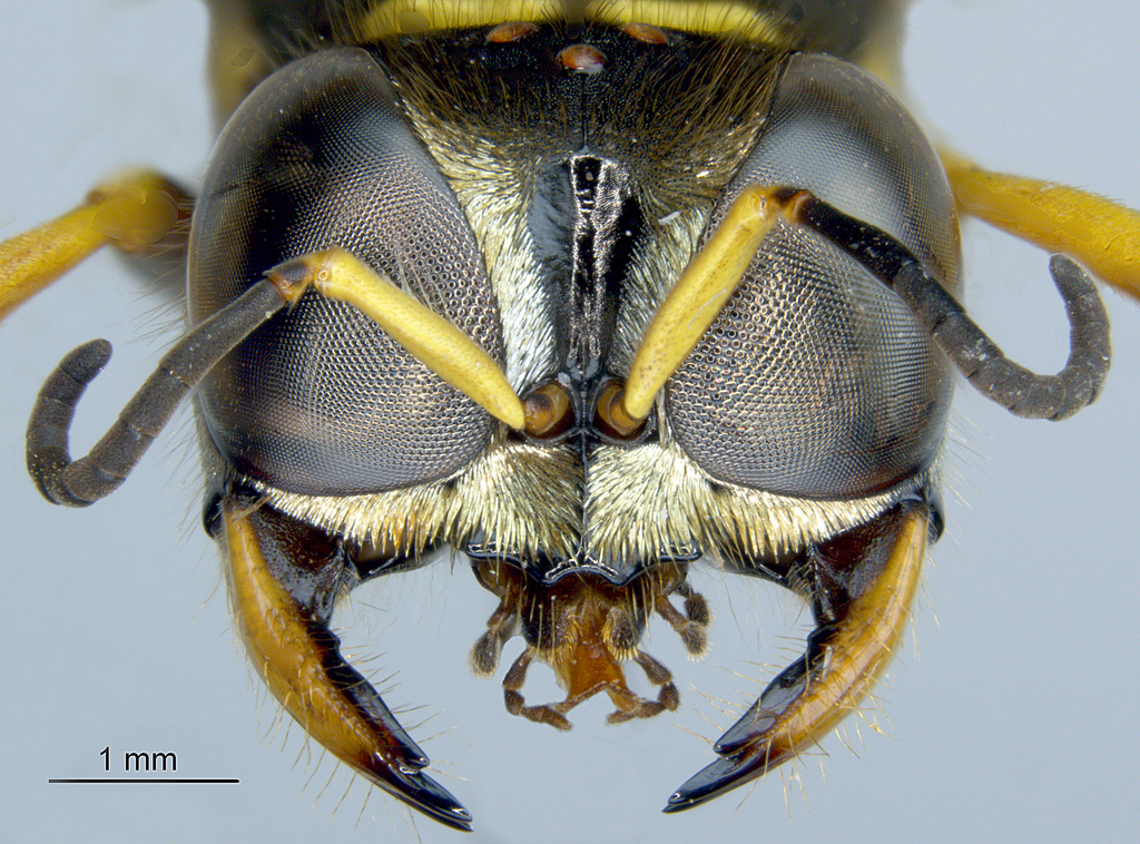
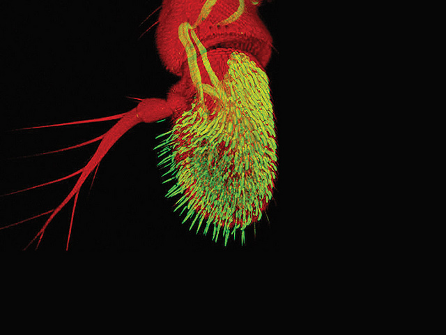
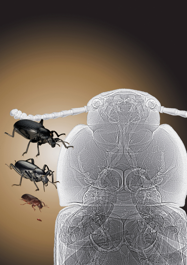
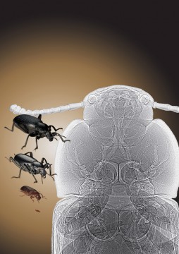
Tracheal tubes are visible in this synchrotron x-ray phase contrast image (right) of a tenebrionid beetle (left). See: Alexander et al., “Increase in tracheal investment with beetle size supports hypothesis of oxygen limitation on insect gigantism,” Proc. Natl. Acad. Sci. USA 104(32), 13198 (August 7, 2007). DOI: 10.1073pnas.0611544104. (Courtesy: The Field Museum of Natural History, Chicago, IL; and Argonne National Laboratory)This image was created with the use of the Advanced Photon Source, located at Argonne National Laboratory, in Illinois. The APS is the source of the brightest X-rays in the Western Hemisphere.Courtesy Argonne National Laboratory.

Comparative thickness of human hair

Credit: Anne Weston, LRI, CRUK. Wellcome Imagesimages@wellcome.ac.ukimages.wellcome.ac.ukThis image shows the difference in thickness of human hair between different ethnic groups. The strand of hair on the left is that of a Caucasian blonde female. The hair on the right is from an Asian male. Hair is normally comprised of three layers, the inner medulla, the cortex and the cuticle. The cuticle is the outermost layer and is comprised of numberous overlapping cells or scales. The cortex makes up the majority of the hair thickness. Interestingly, the inner medulla is not present in blond hair.Scanning electron micrograph

wellcome images – Flickr