
Drosophila Antenna
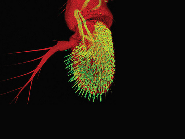
Antennae are paired appendages which arthropods use for sensing. They are the primary olfactory sensors in insects. They are covered with tiny Sensilla which detect chemical signals.
Streptococcus gordonii invading host cells
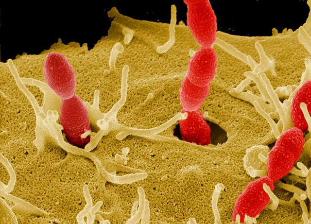
The non-pathogenic non-invasive Streptococcus gordonii (red) is a common and harmless commensal bacterium of the oral human mucosa. By membrane-bound expression of the pathogenicity factor SfbI (Streptococci Fibronectin-binding Protein I) S. goordonii becomes an invasive bacterium.
Image: DSM982 gemini, 5 kV, Inlens:Everhart 50:50
Courtesy of Prof. Dr. Rohde, HZI Braunschweig www.helmholtz-hzi.de/en
Aphids
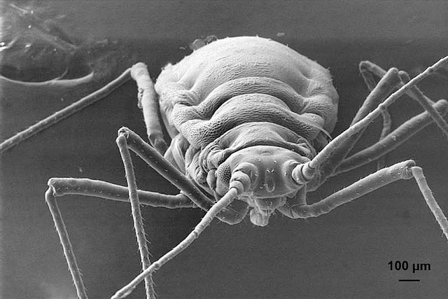
General morphology of an aphid, taken with the EVO® LS 10. The soft body of the insect is not collapsed and details on the surface of the exoskeleton can be seen easily.
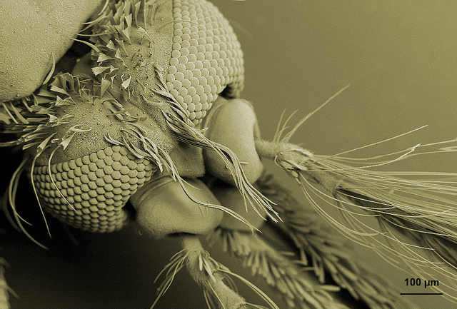
Head and antennae from a male mosquito
Spider
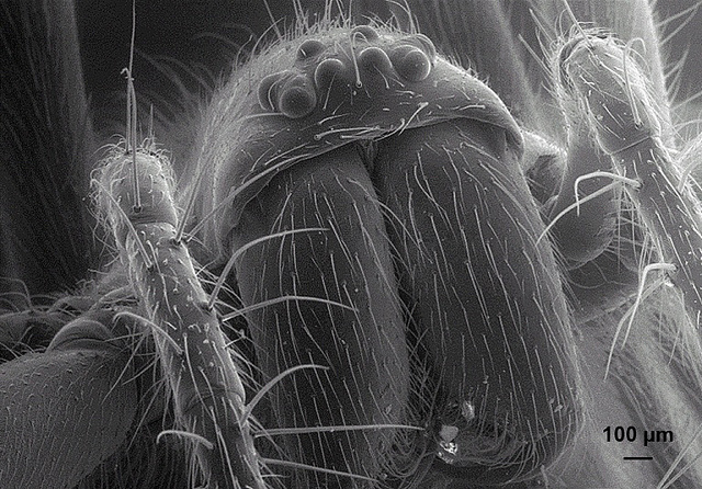
Spider face with detail of the eight eyes, mandibles and body's hair. taken with a EVO® LS 10. The multiple eyes are smooth and rounded without roughness, evidence of absent dehydration.
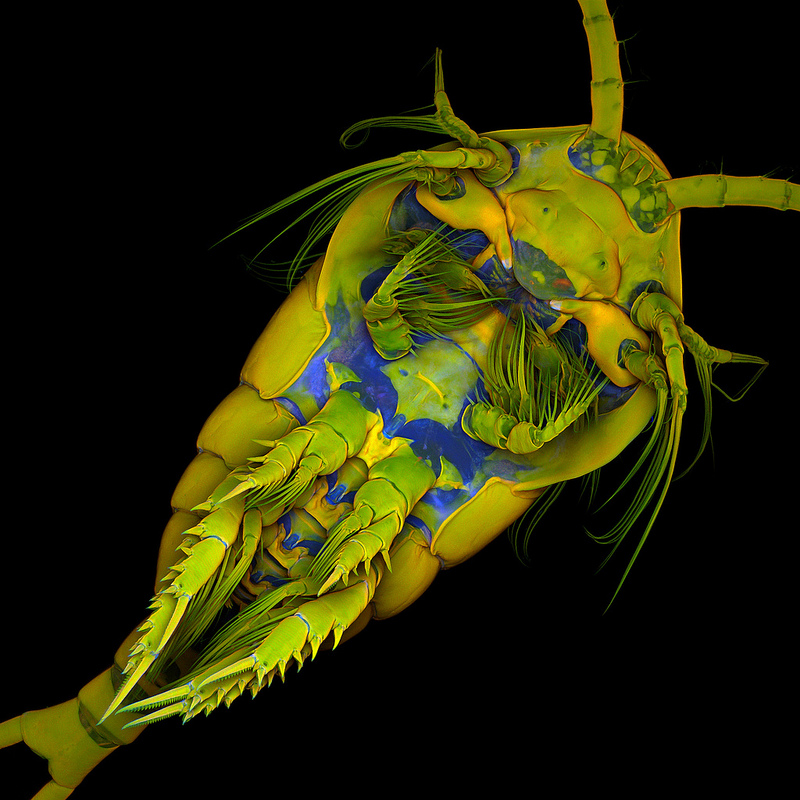
Mite on a millipede leg
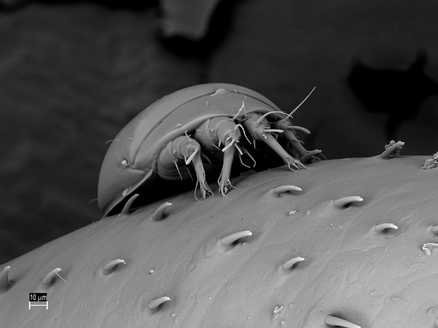
Mite on a millipede leg, imaged with SIGMA (cryo SEM). Plunge frozen into nitrogen “slush” transferred to the SEM at liquid nitrogen temperature, sputter coated with platinum. Sample was provided by the University of Brighton/UK.
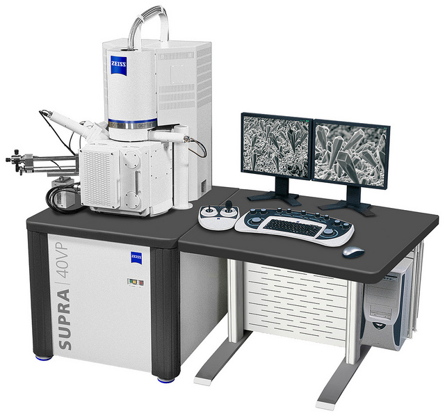
Supra 40 VP
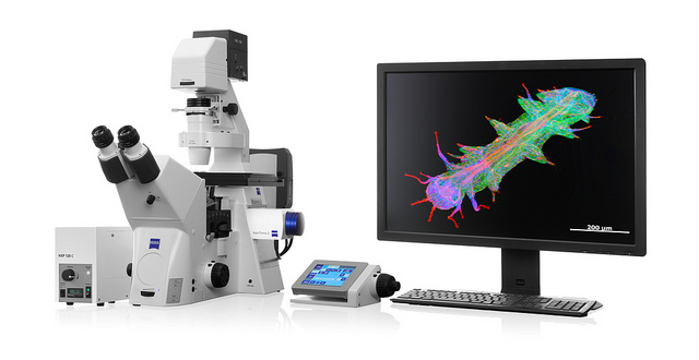
Axio Observer with Apotome.2
Platyneris
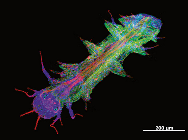
Optical Sectioning image taken using the ZEISS Apotome.2
Platynereis is a genus of marine annelid worm.
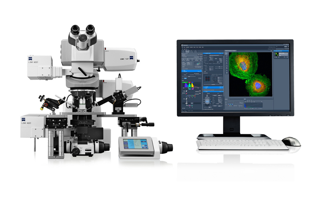
Axio Examiner LSM 7 MP
Ant head
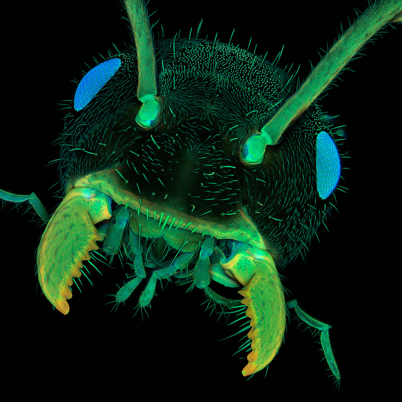
Ant head. Imaged with ZEISS LSM 5 Exciter.Courtesy of Dr. Jan Michels, Zoological Institute, Kiel University
Temora longicornis
Ventral view of a copepod. Imaged with ZEISS LSM 700. en.wikipedia.org/wiki/Copepod
Courtesy of Dr. Jan Michels, Zoological Institute, Kiel University
3D Drosophila Head

Model of an entire Drosophila head. Different sub-structures of the brain and nervous system are color-coded.The raw data for rendering this view was acquired with ZEISS LSMs
Bild: Carl Zeiss Microscopy