
Weitere Unterabschnitte: Biologie | Mikroskopie | Biotechnologie | Chemie | Physik | Physik 2 | Mikrokosmos | Nanokosmos | Wetter und Klima | Weltraum
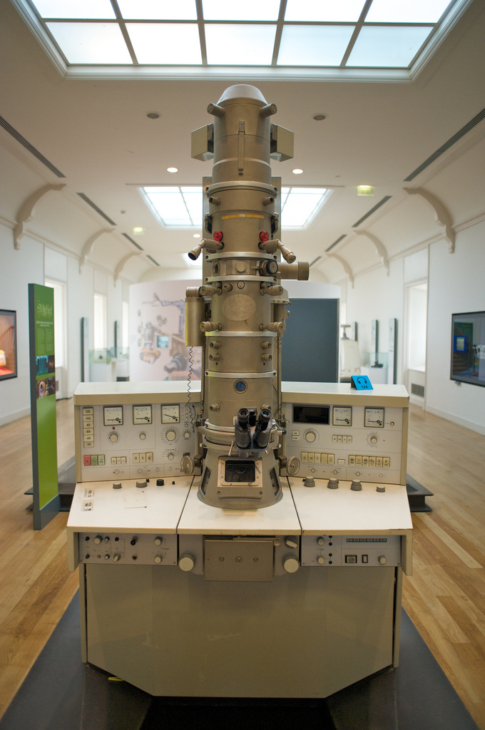
Elektronen-Mikroskopie
David (MK) – Flickr
-
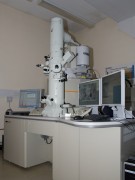
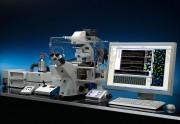
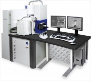
- Electron Microscope in the EMsuite at the OpenUniversity

-
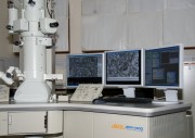
- Electron Microscope in the EMsuite at the OpenUniversity
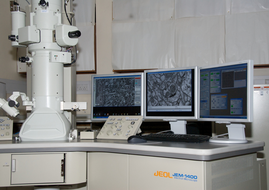
Bild: David (MK)
Arts et Métiers – Siemens Transmission Electron Microscope (TEM)
Carl Zeiss Microscopy – Flickr
-
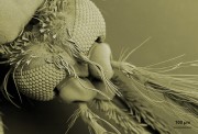
- Drosophila Antenna

-
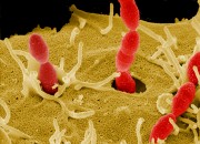
- Streptococcus gordonii invading host cells

-

- Aphids
-
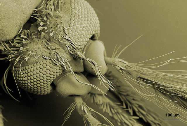
- Head and antennae from a male mosquito
-
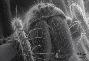
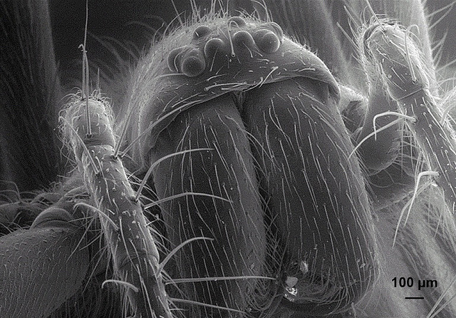
- Spider
-

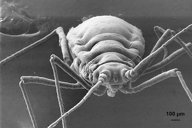
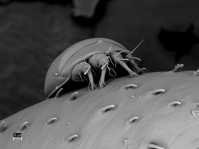
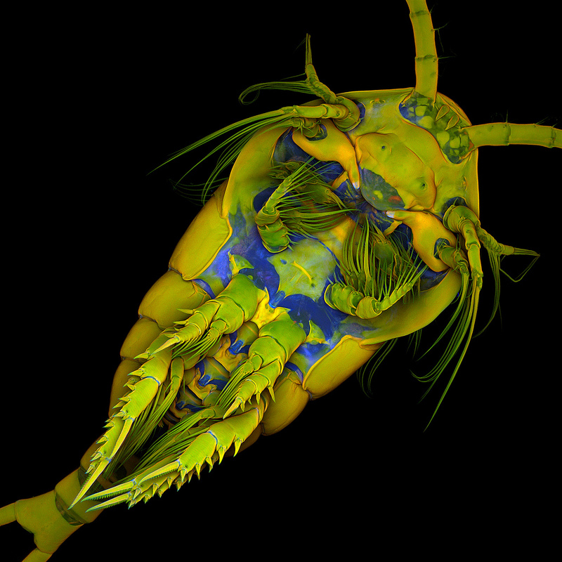
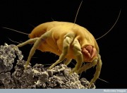
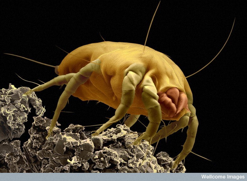
- Mite on a millipede leg
-
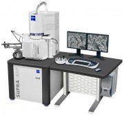
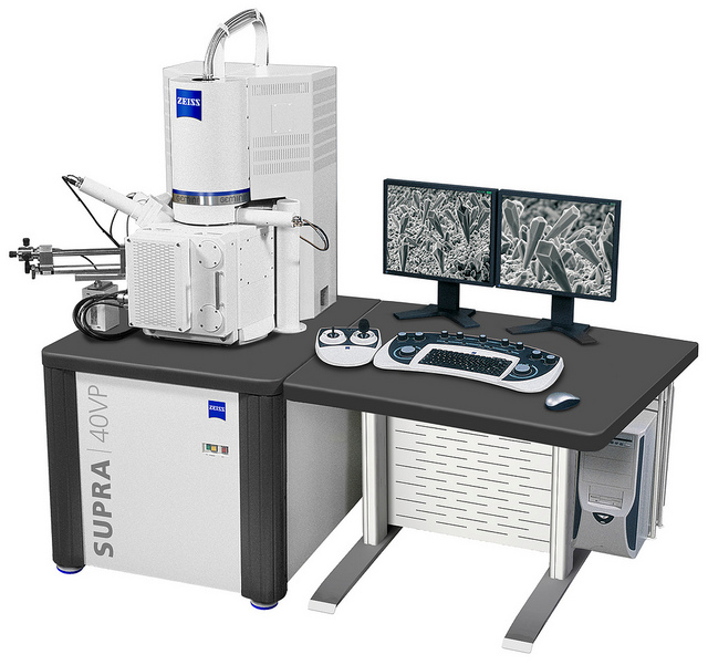
- Supra 40 VP
-
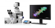
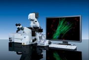
- Axio Observer with Apotome.2
-
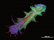
- Platyneris

-

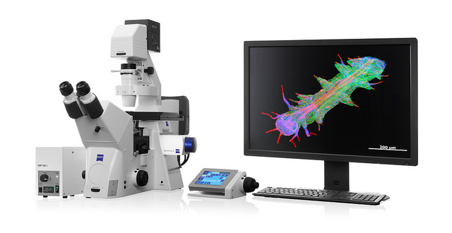
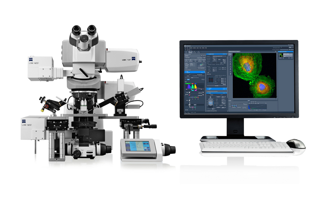
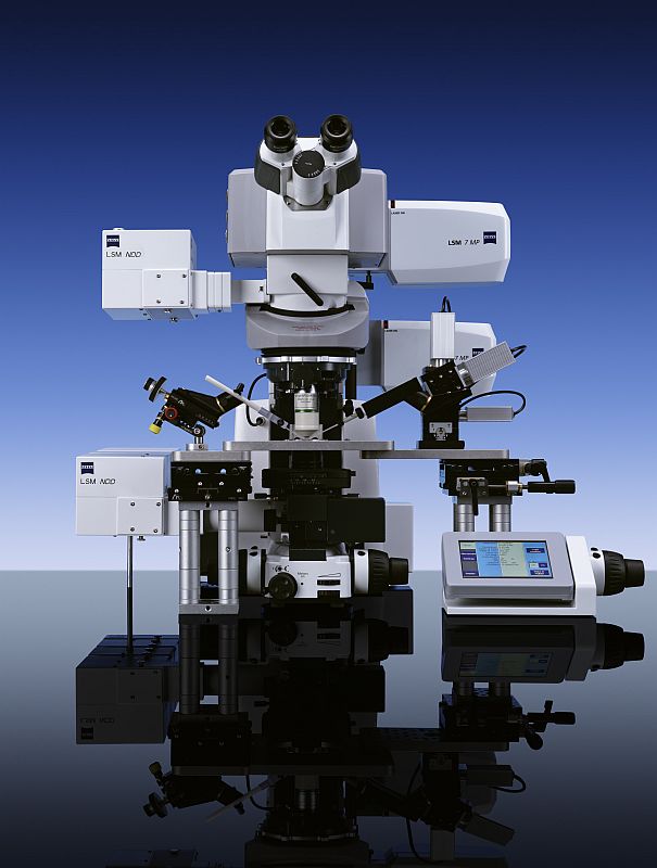
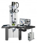
- Axio Examiner LSM 7 MP
-
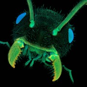
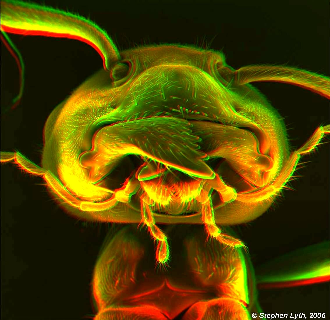
- Ant head
-

- Temora longicornis
-
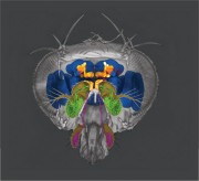
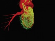
- 3D Drosophila Head

Bild: Carl Zeiss Microscopy
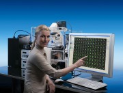
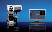
Carl Zeiss AG, Deutschland
-
- Software-Modul AxioVision ASSAYbuilder von Carl Zeiss
-
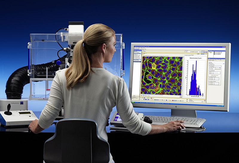
- AxioVision 4.6 – Systemkonfiguration auf Basis des Mikroskops Axio Observer von Carl Zeiss zur Erforschung lebender Zellen.

-
- Mit dem Abbildungssystem Anisotropie Imaging am Laser Scanning Mikroskop LSM 710 von Carl Zeiss können Molekülanordnungen untersucht werden.

-

- Hochauflösende Multiphotonenmikroskopie noch flexibler
-
- Hochauflösung und Flexibilität für die mikroskopische Untersuchung schneller, membrannaher Prozesse

-
- Das Software-Modul AxioVision Physiologie von Carl Zeiss ermöglicht die Kamerabildaufnahme und die Messung physiologischer Parameter in lebenden Zellen sowohl bereits während der mikroskopischen Beobachtung (online) als auch danach (offline).

-
- Neue Mikroskopsoftware verbindet erstmals digitale und molekulare Pathologie

-
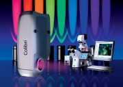
- Colibri, die neuartige Lichtquelle von Carl Zeiss, bietet zahlreiche Vorteile für Fluoreszenzanwendungen in Forschung und Routine.

-
- LIBRA® 120 PLUS

-
- NVision 40 Argon

wellcome images – Flickr
-
- B0008353 Dust mite
-
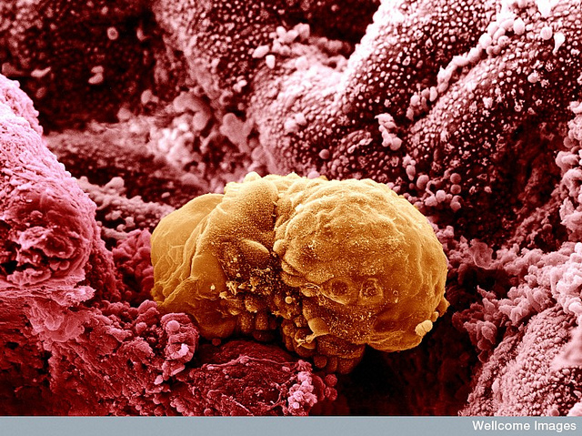
- 6 day old human embryo implanting – coloured
-

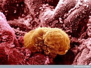
- Human embryonic stem cell
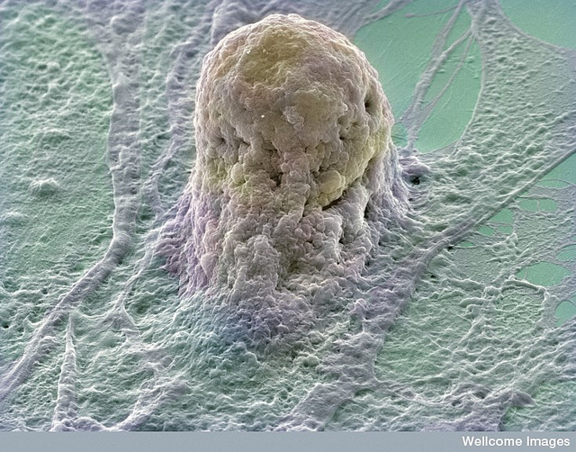
Bild: wellcome images
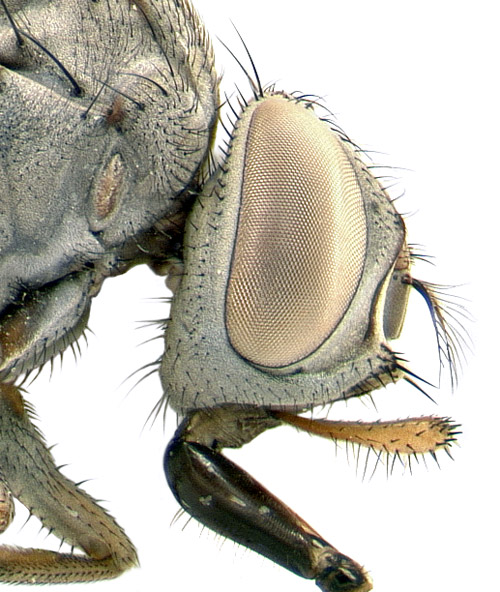
Flies eye
St Stev – Flickr
-
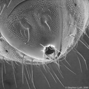
- Stereoscopic Ant
-

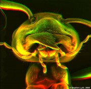
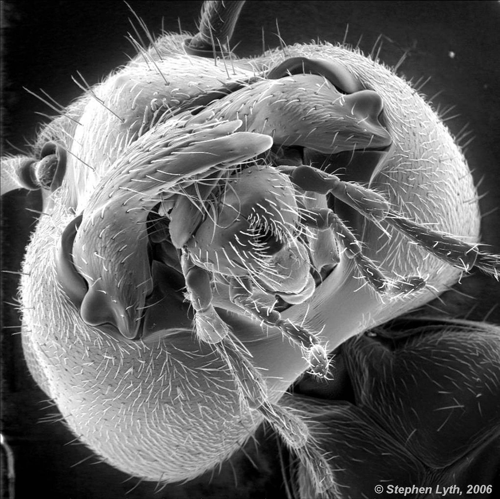
- Ant Portrait
-
- Grotesque Claw

-
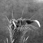
- Ant’s Arse

Bild: St Stev
Beetle tracheas
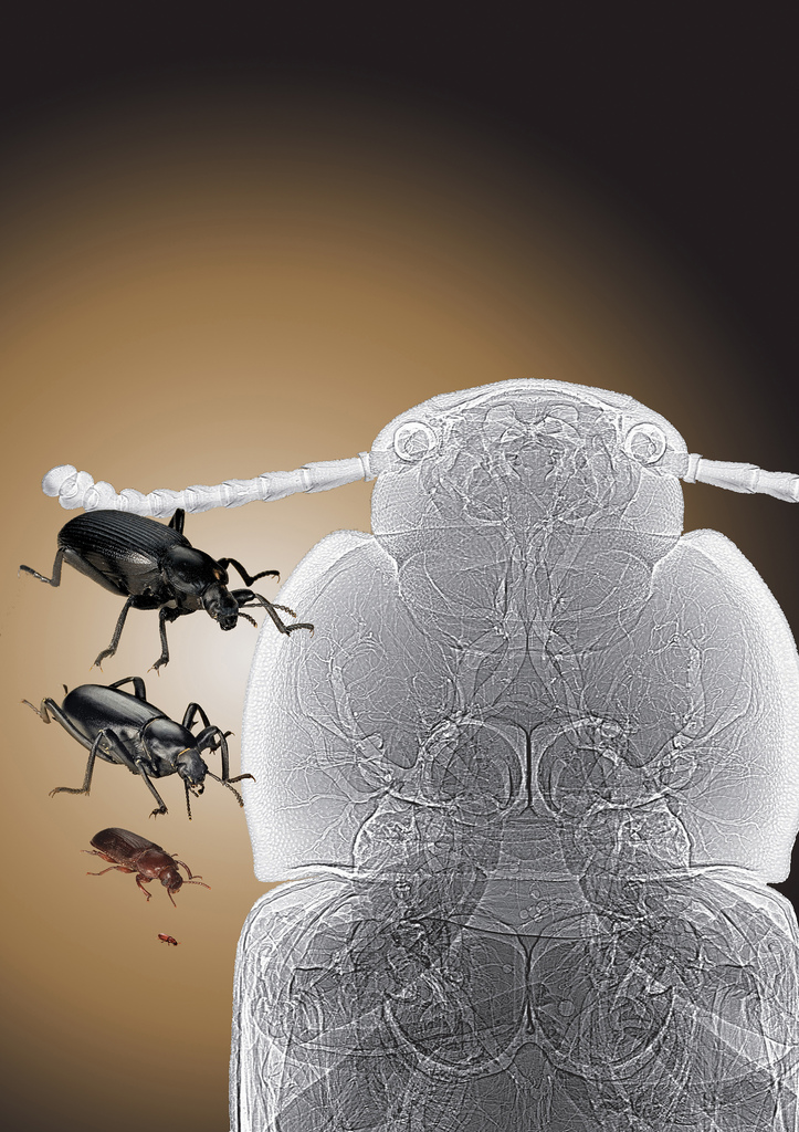
Tracheal tubes are visible in this synchrotron x-ray phase contrast image (right) of a tenebrionid beetle (left). See: Alexander et al., “Increase in tracheal investment with beetle size supports hypothesis of oxygen limitation on insect gigantism,” Proc. Natl. Acad. Sci. USA 104(32), 13198 (August 7, 2007). DOI: 10.1073pnas.0611544104. (Courtesy: The Field Museum of Natural History, Chicago, IL; and Argonne National Laboratory)This image was created with the use of the Advanced Photon Source, located at Argonne National Laboratory, in Illinois. The APS is the source of the brightest X-rays in the Western Hemisphere.Courtesy Argonne National Laboratory.
Comparative thickness of human hair

Credit: Anne Weston, LRI, CRUK. Wellcome Imagesimages@wellcome.ac.ukimages.wellcome.ac.ukThis image shows the difference in thickness of human hair between different ethnic groups. The strand of hair on the left is that of a Caucasian blonde female. The hair on the right is from an Asian male. Hair is normally comprised of three layers, the inner medulla, the cortex and the cuticle. The cuticle is the outermost layer and is comprised of numberous overlapping cells or scales. The cortex makes up the majority of the hair thickness. Interestingly, the inner medulla is not present in blond hair.Scanning electron micrograph
Bild: wellcome images
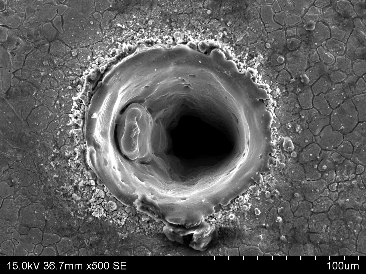
Hole drilled in stainless steel
wellcome images – Flickr
-
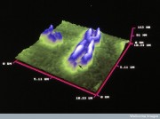
- Scanning probe image of chromosome 1
-
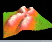
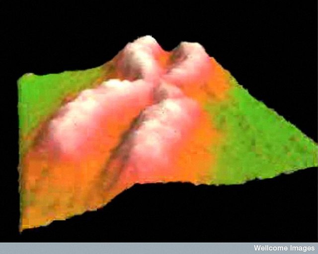
- Fragile X chromosome, atomic force microscope

Bild: wellcome images