
IBM NanoMRI closeup
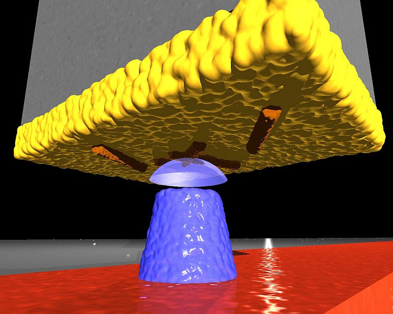
An artistic view of the magnetic tip (blue) interacting with the virus particles at the end of the cantilever.
IBM NanoMRI
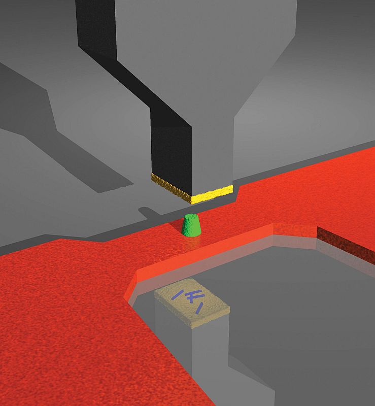
Rendering of the key elements of a magnetic resonance force microscope. An ultrasensitive silicon cantilever detects the tiny magnetic force between a nanoscale magnetic tip (green) and the hydrogen nuclei present in the virus particles placed at the end of the cantilever (blue, seen in the reflection). Nanoscale magnetic resonance imaging is achieved by manipulating the hydrogen nuclei in the sample with a radiofrequency magnetic field generated by a "microwire" (red). A sensitivity improvement of 100 million is achieved compared to conventional magnetic resonance imaging.