Medical Imaging
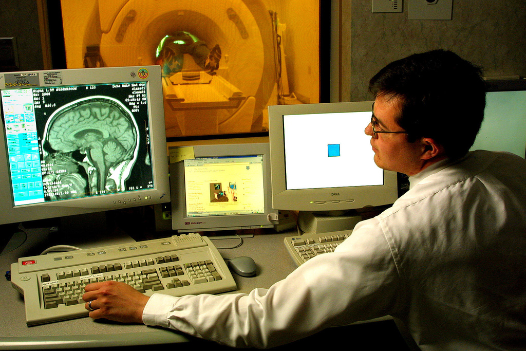
Medical Augmented Reality: Easy Visualization In-Situ
12 Week Sonar Scan
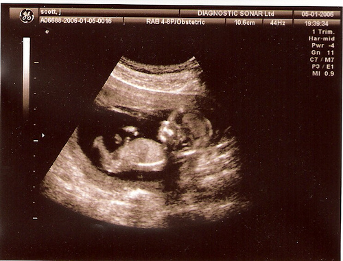
Bild: Jo and Paul's pics
MRT (Magnetresonanztomographie)
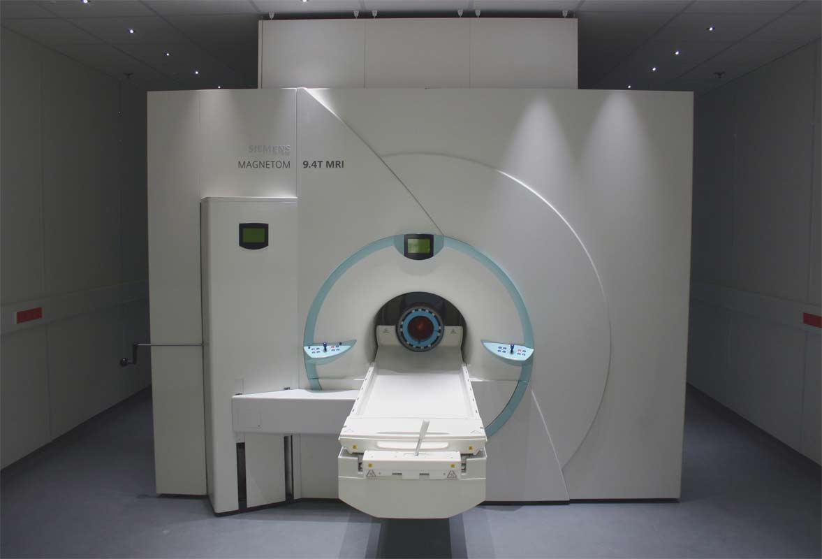
Einsichten in den Körper: Mit den drei Tomografen des Magnetresonanzzentrums untersuchen Wissenschaftler Menschen und Tiere. Hier zu sehen ist der 9,4-Tesla-Magnet mit einer Liegefläche für die Probanden.
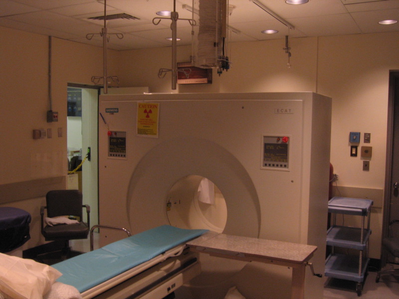
PET – Positron Emission Tomography scanner #2
B1SHOP – Flickr
-
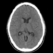
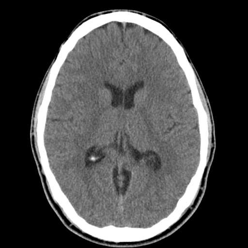
- Brain Scan: Top 02
-
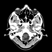
- Brain Scan: Top 01

-

- Brain Scan: side
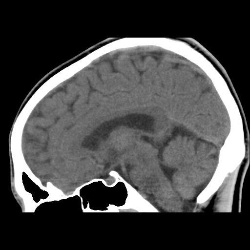
Bild: B1SHOP
MRT (Magnetresonanztomographie)
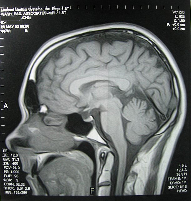
Bild: jsmjr
spontaneous pneumothorax (Röntgenaufnahme)
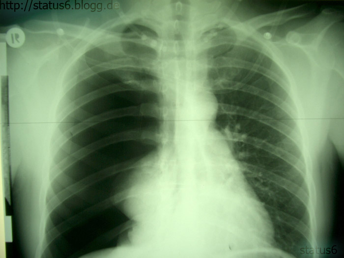
Bild: status6
MRT (Magnetresonanztomographie)

Bild: pixieclipx
X-ray of the foot of a patient

Canadian Forces Medical Officer Major Max Talbot checks the X-ray of the foot of a patient at the Role 3 medical facility at Kandahar Airfield, Afghanistan. Major Talbot is currently serving with the Canadian Forces' Joint Task Force Afghanistan.
Bild: lafrancevi
chiari_mri

An MRI shot of my brain before my posterior fossa decompression (brain surgery) for Arnold Chiari Malformation.
See the white blob towards the bottom right? That's not supposed to be there - it's my cerebellum sinking down into my spinal column. That's what Chiari does. The brain drops down & blocks the flow of CSF (spinal fluid.) Bad news & I was sick for almost three years before getting diagnosed in the fall of 2005.
Posterior fossa decompression is where they remove part of the back of your skull & create a patch that holds your cerebellum up.
Bild: chaserpaul
Funktionelle Magnetresonanztomographie – Wikipedia
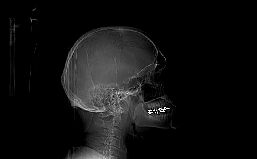
Röntgenaufnahme der Halswirbelsäule
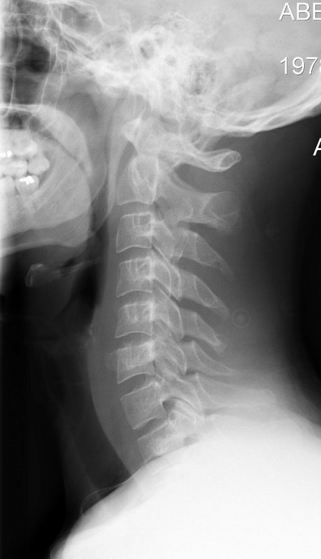
Bild: Andrew Ciscel
Activation of Brain Region Predicts Altruism.
Important Note: this image is not mine but was available for download, and is the property of:
Scott Huettel
Digital Image IMG0031
File 0332/02
© Duke University Photography Jim Wallace
Bild: --Tico--