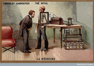
Wilhelm Conrad Roentgen looking into an X-ray screen

Credit: Wellcome Library, London. Wellcome Images images@wellcome.ac.uk images.wellcome.ac.uk Wilhelm Conrad Roentgen looking into an X-ray screen placed in front of a man's body and seeing the ribs and the bones of the arm. Chromolithograph. Chromolithograph Published: [1896/1900]

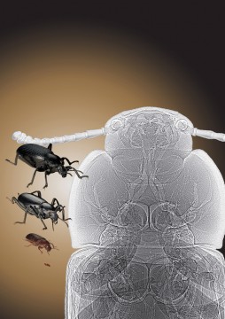
Beetle tracheas

Tracheal tubes are visible in this synchrotron x-ray phase contrast image (right) of a tenebrionid beetle (left). See: Alexander et al., “Increase in tracheal investment with beetle size supports hypothesis of oxygen limitation on insect gigantism,” Proc. Natl. Acad. Sci. USA 104(32), 13198 (August 7, 2007). DOI: 10.1073pnas.0611544104. (Courtesy: The Field Museum of Natural History, Chicago, IL; and Argonne National Laboratory)This image was created with the use of the Advanced Photon Source, located at Argonne National Laboratory, in Illinois. The APS is the source of the brightest X-rays in the Western Hemisphere.Courtesy Argonne National Laboratory.
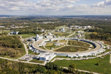
Advanced Photon Source

Based on any of a variety of different criteria ? number of users, publications or deposits in the Protein Data Bank, for example ? the Advanced Photon Source at Argonne National Laboratory stands out as the premier hard X-ray research facility in the Western Hemisphere.

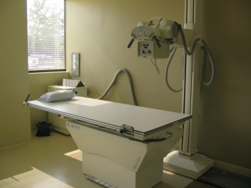
Röntgen im digitalen Zeitalter – Flachdetektoren verbessern Workflow und Diagnosen

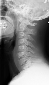
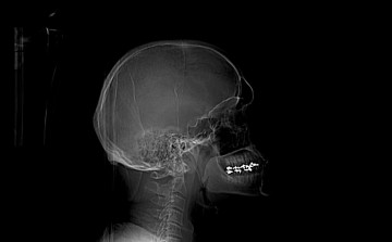
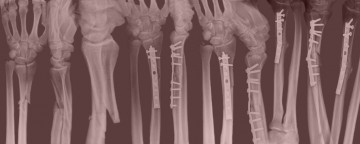
Montag, 05.12.2005 Röntgen ist das älteste bildgebende Verfahren in der Medizin. Am Vorgehen hat sich über hundert Jahre hinweg nicht viel geändert: Skelett- oder Lungenaufnahmen wurden auf Film belichtet und vor einem Leuchtschirm analysiert. Die Fotographie war Ende der 90er Jahre längst im digitalen Zeitalter angekommen, als die digitale Bildverarbeitung über die Flachdetektoren ihren Einzug in die Praxen und Kliniken auf dem Gebiet der Radiographie mit Echtzeitbilddarstellung antrat.