
Weitere Unterabschnitte: Übersicht | Diagnose | Therapie
Diagnose
Magnetresonanz (MR)
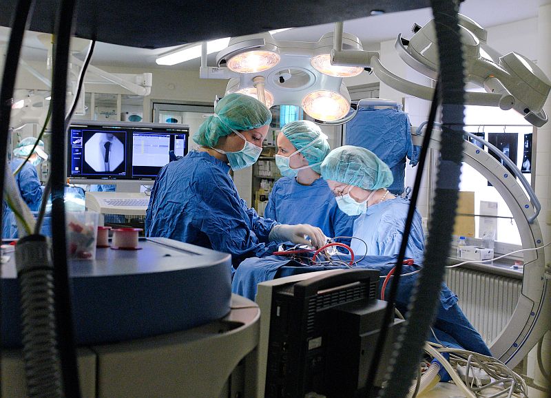
Siemens AG – Hightech-Medizin jetzt auch für Haustiere
-
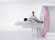
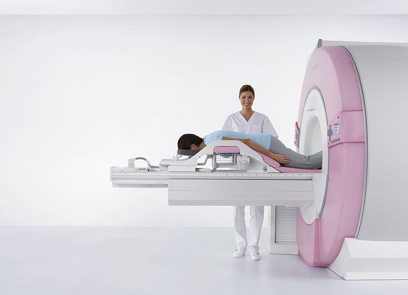
- Innovation in der Frauenheilkunde %u2013 der neue MR-Brustscanner von Siemens
-
- Innovation in der Frauenheilkunde – der neue MR-Brustscanner von Siemens
-

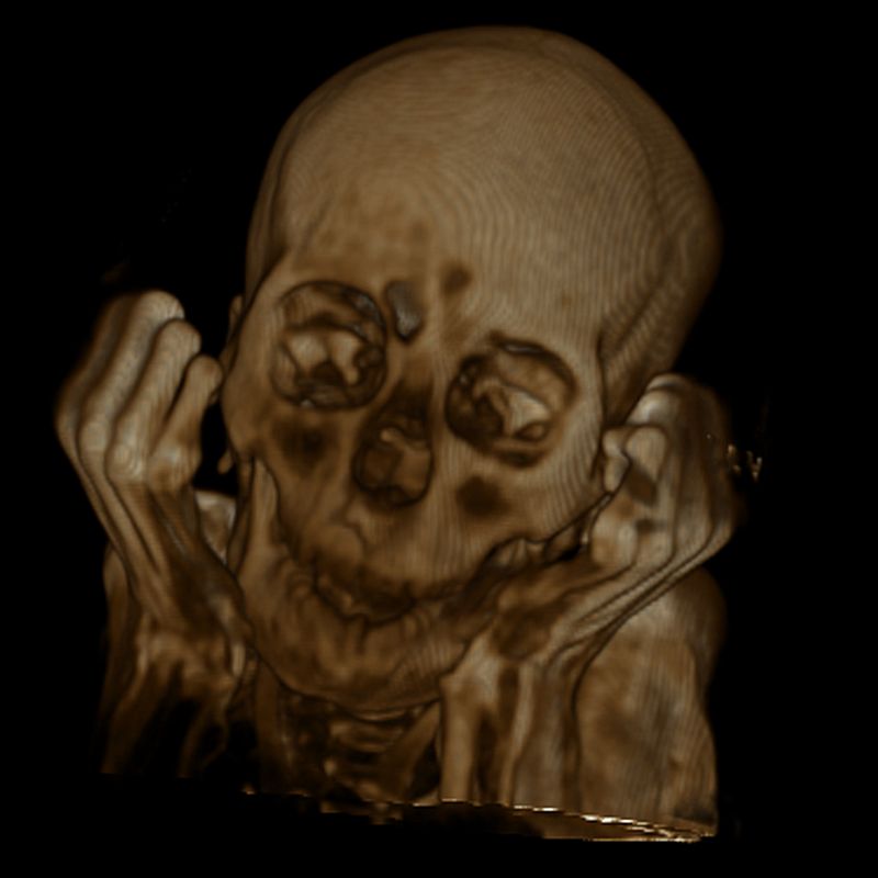
- Mumientest zeigt Magnetresonanz-Röhre in Höchstform:
-
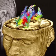
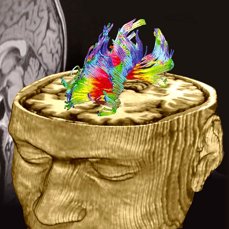
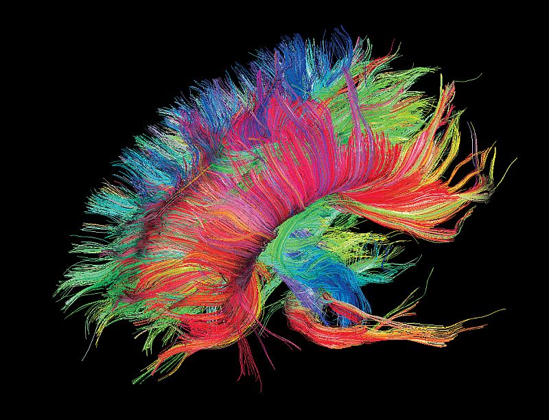
- Nerven im Gehirn
-
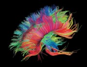
- Nerven im Gehirn
-
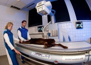
- Hightech-Medizin jetzt auch für Haustiere

-

- Hightech-Medizin jetzt auch für Haustiere
-
- Hightech-Medizin jetzt auch für Haustiere
-

- Hightech-Medizin jetzt auch für Haustiere
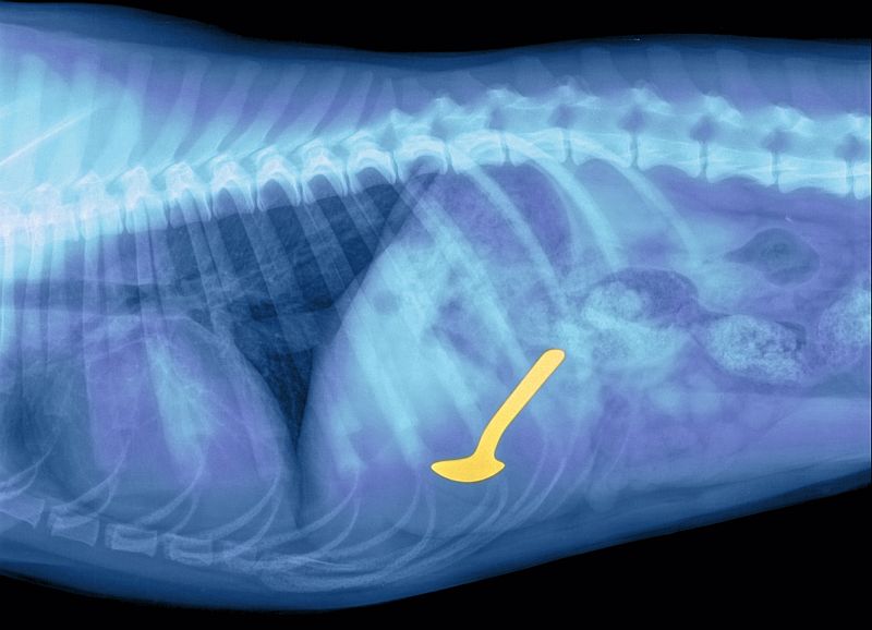
Foto: Siemens AG
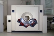
Das neue Magnetresonanzzentrum am Max-Planck-Institut für biologische Kybernetik hat am 12. Juli 2007 seinen Betrieb aufgenommen
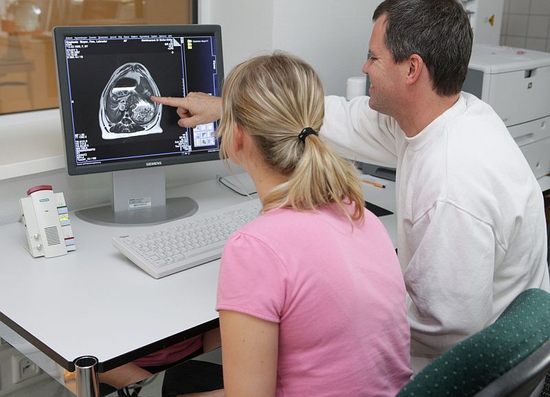
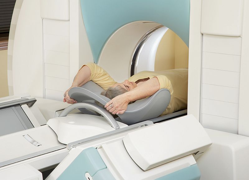
MRT (Magnetresonanztomographie)
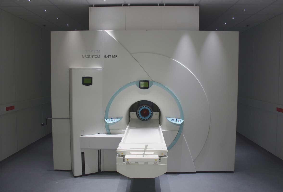
Einsichten in den Körper: Mit den drei Tomografen des Magnetresonanzzentrums untersuchen Wissenschaftler Menschen und Tiere. Hier zu sehen ist der 9,4-Tesla-Magnet mit einer Liegefläche für die Probanden.
MRT (Magnetresonanztomographie)

Bild: pixieclipx
Funktionelle Magnetresonanztomographie – Wikipedia
fMRI – Functional magnetic resonance imaging scanner
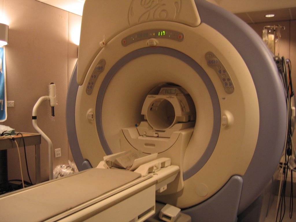
Paying another visit to a friend's office. I found that the fMRI was not in use so I got permission to take a few pics. Since it is an fMRI rather than the older MRI, it is a bit more compact
Bild: MacRonin47
chiari_mri

An MRI shot of my brain before my posterior fossa decompression (brain surgery) for Arnold Chiari Malformation.
See the white blob towards the bottom right? That's not supposed to be there - it's my cerebellum sinking down into my spinal column. That's what Chiari does. The brain drops down & blocks the flow of CSF (spinal fluid.) Bad news & I was sick for almost three years before getting diagnosed in the fall of 2005.
Posterior fossa decompression is where they remove part of the back of your skull & create a patch that holds your cerebellum up.
Bild: chaserpaul
MRT (Magnetresonanztomographie)
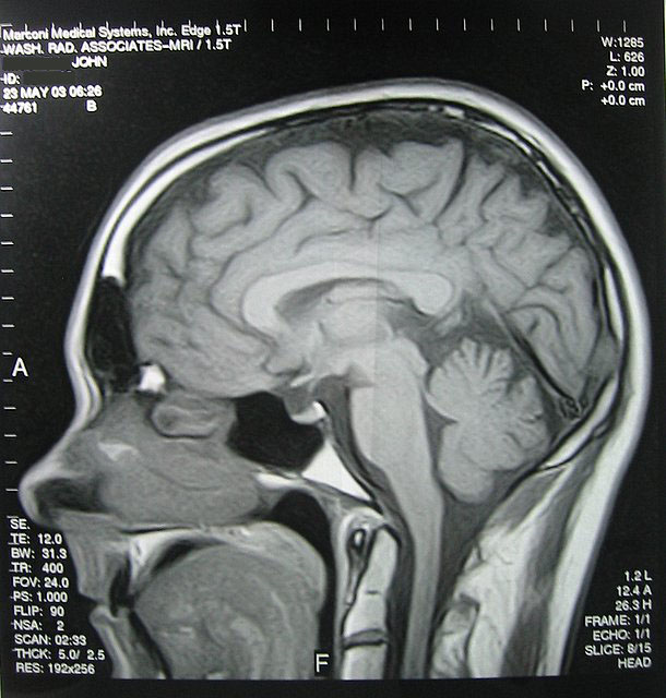
Bild: jsmjr
B1SHOP – Flickr
-
- Brain Scan: Top 02
-
- Brain Scan: Top 01

-

- Brain Scan: side
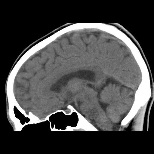
Bild: B1SHOP
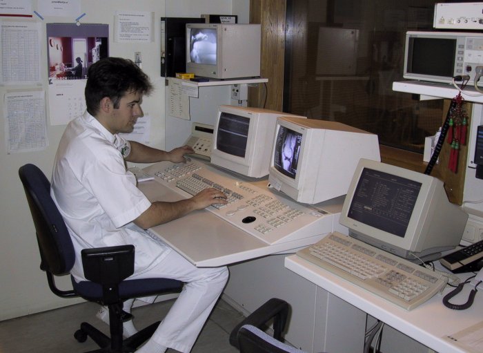
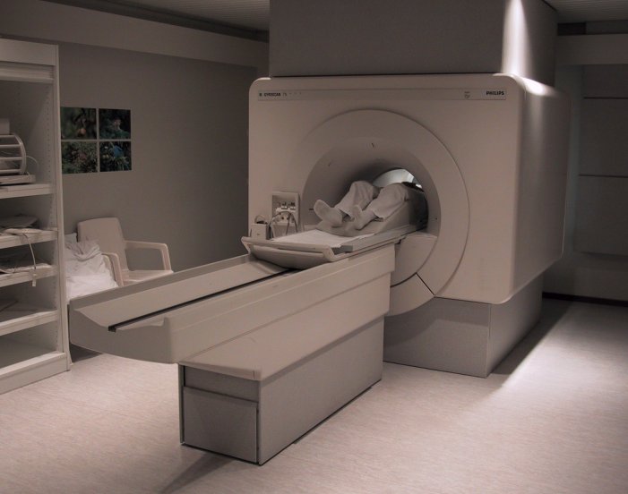
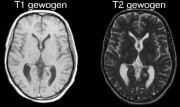
Neurochirurgie Zwolle: Magnetresonanz
-

- MRI_MRIconsole.jpeg
-

- MRI_MRIscanner.jpeg
-
- MRI_normaleMRI.jpeg

-
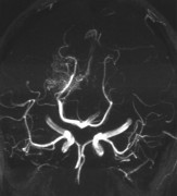
- MRI_MRA.jpeg

Nuklearmedizin
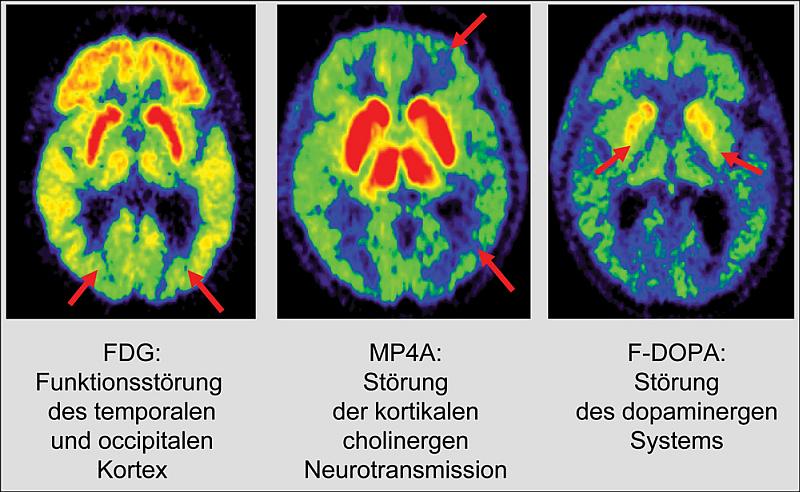
PET scan of an healthy brain compared to a brain at an early stage of Alzheimer’s disease.
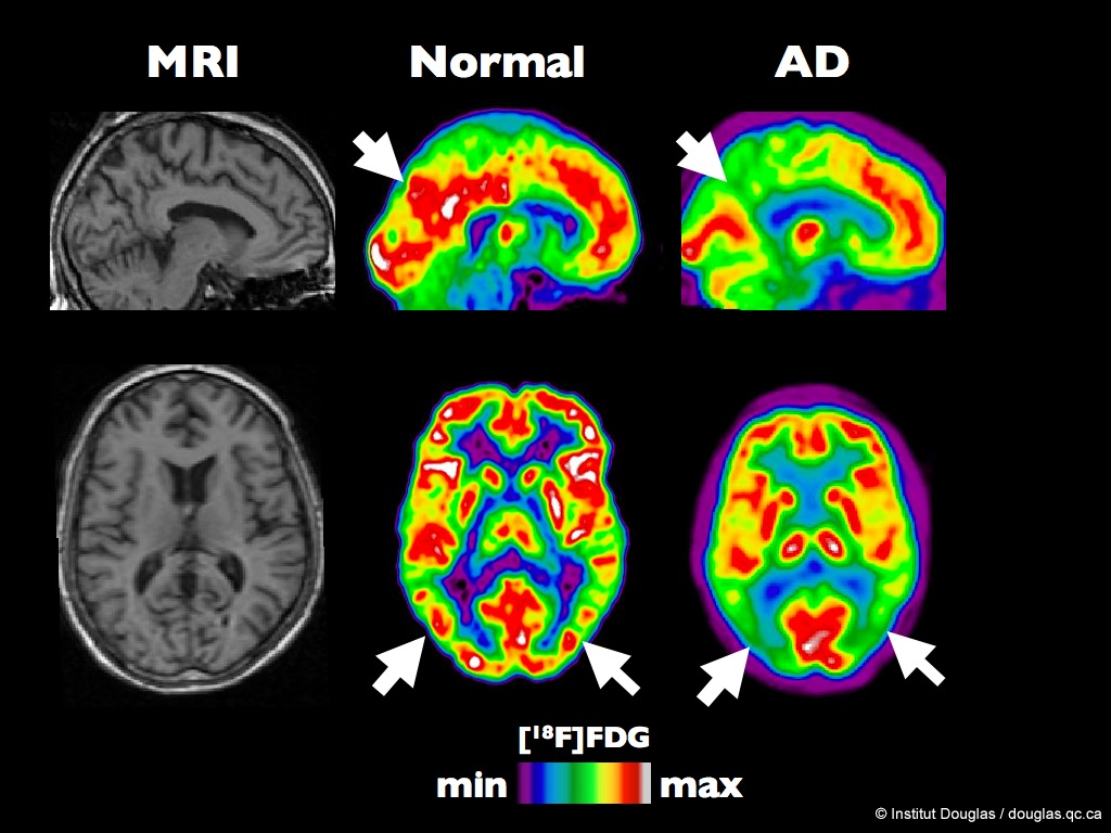
The figure shows MRI, and typical [18F]FDG images from a healthy volunteer and a Alzheimer Disease (AD) patient. The color scale express the rate of glucose utilization (from minimum to maximum). In these PET (Positron Emission Tomography) images (column 2 and 3), the white and red colors represent the regions of the brain with the highest glucose utilization rates while the green and blue represent the hypometabolic brain areas.
The arrows show the posterior temporo-parietal junction (lower row) and the posterior cingulate gyrus (top row).
Bild: Institut Douglas
Brookhaven National Laboratory – Flickr
-

- Pre-PET Headgear (Positron Emission Tomography)
-
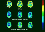
- Cocaine brain scans, PET

Neue Technologie von Siemens für Gamma-Kamera in der molekularen Bildgebung
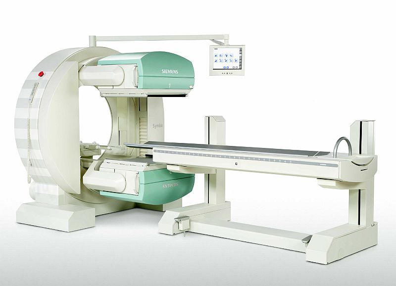
17.04.2008 | Symbia E enthält modernste SPECT-Technologie und ist mit verbesserter Elektronik, neuem Design sowie einer überarbeiteten Chassis ausgestattet. Sie verfügt gleichzeitig über alle Vorteile der am Markt etablierten Gamma-Kamera e.cam von Siemens, von der weltweit mehr als 4000 in über 120 Ländern installiert sind. Symbia E ist in der Lage, aufgrund ihrer hohen Bildqualität und Zuverlässigkeit den Workflow der Anwender zu beschleunigen.
Siemens AG – Pressebilder
-
- IQ SPECT: Intelligente Funktion für Gammakamera beschleunigt nuklearmedizinische Untersuchungen

-
- IQ SPECT: Intelligente Funktion für Gammakamera beschleunigt nuklearmedizinische Untersuchungen
Max Planck Gesellschaft – Geschärfter Blick ins kranke Gehirn
-
- Der neue Positronen-Emissions-Tomograph für das Gehirn. Der Bildausschnitt zeigt die Detektorköpfe.

-
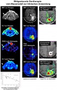
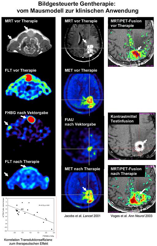
- Bildgesteuerte Gentherapie
-
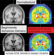
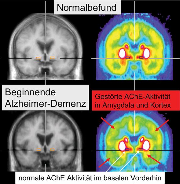
- Nachweis der Störung des cholinergen Systems in Amygdala und Kortex mit einer speziellen Markierungssubstanz und PET bei beginnender Alzheimer-Demenz.
-
- PET-Aufnahme mit verschiedenen Markierungssubstanzen (Multitracer-PET) bei Lewy-Körperchen-Demenz.
Mit freundlicher Genehmigung Max-Planck-Gesellschaft
First Class Health Care in Panama
Panama is not just pretty - it's modern. Top-notch health care is available at much lower rates than most other countries. Highly trained doctors with superb patient care.
Bild: thinkpanama